
Dengue, caused by a mosquito-borne virus, is a serious global health concern. It is prevalent in tropical and sub-tropical areas like southeast Asia, America and western pacific regions. Early diagnosis (identification) of such diseases is essential to provide timely management to the patient and prevent health deterioration to severe illness.
However, existing diagnostic methods like virus isolation, nucleic acid detection are time-taking (may even take a week), require a lot of expensive equipment and reagents and need technical expertise, limiting their use for early detection. The need of the hour is a rapid, inexpensive, user-friendly on-site diagnosis that requires minimal infrastructure and is scalable to the community level.
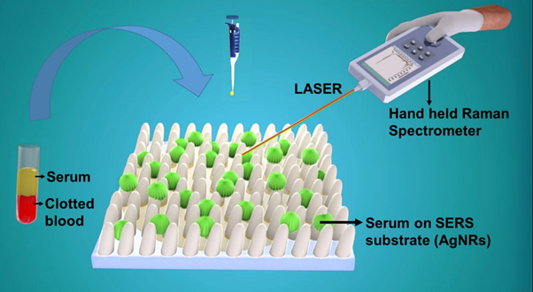
The technique presented by the GLAD research group at IIT Delhi’s Physics Department meets most of these requirements. They have created a Surface Enhanced Raman Spectroscopy based Platform (SERS) that gives dengue tests results within an hour.
The main idea behind the set-up is the well-known technique of Raman Spectroscopy.
Raman Spectroscopy
A sample scatters only a tiny part of light falling on it, about 1 %. 99% of this scattered light consists of photons of the same frequency as the incident light known as Rayleigh-scattering. The rest of it is shifted in both wavelength and energy, known as Raman-scattered light.
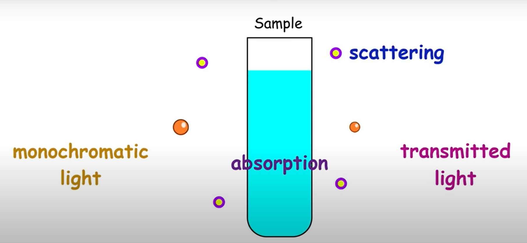
The spectrum of the Raman-scattered light depends on the molecular constituents of the sample and hence is specific to that material. That makes it helpful to identify any substance (including biological materials) by its Raman spectrum. In fact, they are known as molecular fingerprints for this reason.
A popular method for detecting viruses in the body is to check for the presence of their antigens. An antigen is a toxin or other substance given off by a virus that causes an immune response in our body. Raman Spectroscopy serves as a great tool to detect and identify these antigens. For example, the presence of NS1 antigen confirms Dengue.
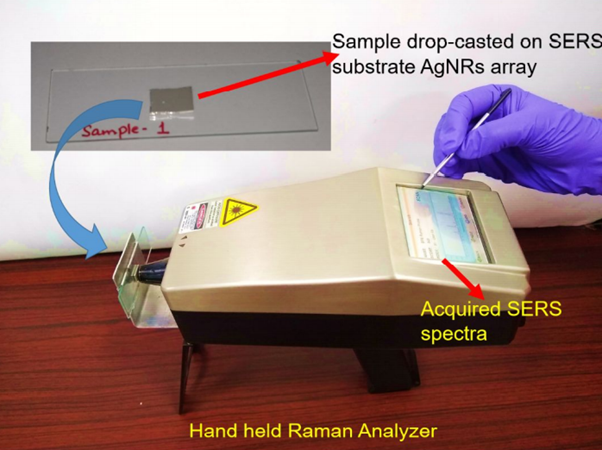
This can be quickly done on-site at a hospital/institute with the help of a handheld Raman spectrometer. It shoots a laser beam at the sample, collects the scattered light and displays the resulting spectrum (intensity – wavelength graph).
However, biological materials scatter light poorly, further reducing the already low scattered light intensity, leading to weak Raman signals that make it difficult to detect the antigen at lower concentrations.
SERS Substrate
To tackle this problem, , the research group headed by Principal Investigator Professor J.P. Singh used a technique called GLAD to fabricate thin roughened up metal surfaces (usually made of noble metals like Gold) with different shapes, built with precision up to the nanometer range.
Then, silver nanoparticles (having a diameter between 1 to 100 nm) from a solution were deposited on the metal surface.
The resulting metal surface, known as a SERS platform, acts as an amplifier, i.e. when shone on with a laser, the surface electrons on these metal structures get excited and create an enhanced electric field localized near the surface.
The degree of enhancement greatly depends on the dimensions and shape of the SERS platform and the nature of the nanoparticles used. However, by utilizing the optimum combination, the researchers obtained electric fields as high as 108 times the original one.
When the dengue samples to be tested were adsorbed on the platform, and their spectra analyzed, the Raman signals obtained were enhanced by the same factor!
With this method, spectral data can be obtained even at low concentrations of the antigen. This increases the specificity (proportion of true negatives) of the diagnostic test by eliminating false negatives. The use of SERS would tremendously increase the application of Raman Spectroscopy in medicine and allied fields.
Testing
The handheld device has been successfully tested on clinical blood samples collected from a fever clinic at the Indian Council Of Medical Research (ICMR) – NIMR, New Delhi.
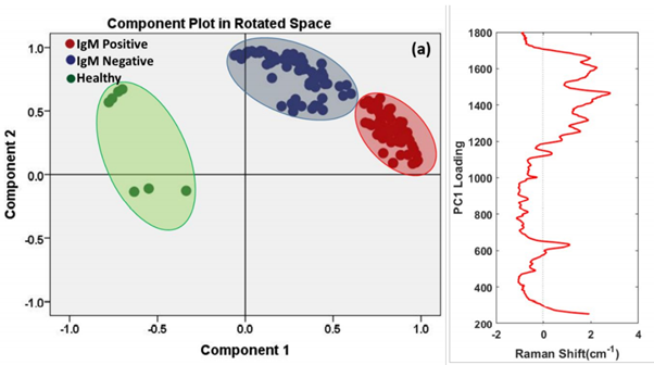
Blood samples were taken from fever patients as well as healthy individuals, and the spectral data obtained was fed into a Principal Component Analyzer (PCA) that is a statistical tool for data analysis. Based on the input data, It accurately differentiated between the three sets of blood samples, Dengue positive, Dengue negative and healthy patients for any samples tested from thereon.
The SERS technique is not only limited to Dengue and can be extended to detect a host of other viruses and bacteria. For example, in collaboration with the ICMR- National Aids Research Institute (NARI), the research group successfully demonstrated the detection of the human immunodeficiency virus (HIV-1) through this handheld SERS platform.
Conclusion
It may seem that sophisticated fabrication methods drive up the expenses, however, a SERS chip developed by this technique costs no more than a dollar, and the Raman spectrometer makes for a one-time investment by the hospital/institute. In all, the SERS platform shows excellent potential to replace the traditional diagnostic methods used nowadays.