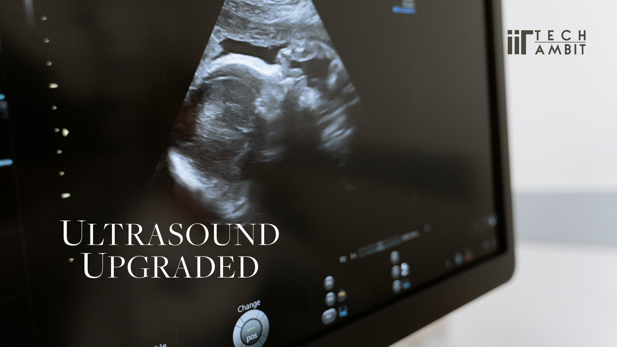
The introduction of ultrasound in the late 1950s created a revolution in the field of medical imaging. Since then, ultrasound technology has proved to be a boon to the medical industry owing to its advantages such as non-invasiveness, portability, safety, and ease of imaging with minimal strain in the patients. It uses high frequency non-ionising sound waves to create images and find applications in Sonar (sound navigation and ranging), non-destructive testing of materials, medical diagnostics, and therapeutics. Medical ultrasound imaging, commonly termed ultrasonography, is often used in cardiology, gynaecology, abdominal imaging etc. to help diagnose and detect abnormalities or to provide visual assistance with surgeries and biopsies.
With the advancements in the field of electronics and computers, ultrasound imaging is evolving as a miniaturized, portable, and more affordable modality that provides exceptional image quality and performance. But studies show that more research is required to develop cost effective methods for ambulatory care to ensure that evidence-based treatments are initiated and treatment goals are attained and maintained over time. This emphasizes the necessity for an affordable and portable diagnostic system for ambulatory scenarios. In this regard, there have been various efforts towards developing portable ‘ultra-fast’ ultrasound imaging systems for point of care and bedside applications. One such effort is a research project being worked on by Dr. Mahesh and Mr. Madhavanunni from IIT Palakkad.
Dr. Mahesh Raveendranatha Panicker is an Assistant professor in the Electrical Engineering department, IIT Palakkad. Dr. Mahesh received his Ph.D. from the School of Computer Engineering, Nanyang Technological University, Singapore in 2009. He is a senior member in the Institute of Electrical and Electronics Engineers (IEEE) and is also a Six Sigma Green Belt certified TRIZ practitioner. His research interests include signal processing and analytics, diagnostic ultrasound imaging, biomedical circuits and systems, industrial prognostics and health monitoring. One of his recent and important contributions in the area of diagnostic ultrasound imaging is the development of the Automated Lung Ultrasound analysis tool. The team was led by Dr. Mahesh and you can read more about the tool in the article written by Varun Ganta.
Madhavanunni A N is a graduate student pursuing his Ph.D. in the area of Biomedical Ultrasound Imaging under the guidance of Dr. Mahesh at IIT Palakkad. Dr. Mahesh and Mr. Madhavanunni started working on an ultrasound imaging system that is both portable and affordable and gives better results than the existing portable ultrasound systems. The system being developed by this team is a high frame rate portable flow imaging system. These systems have a very limited commercial availability.
How does it work?
A typical ultrasound imaging system mainly consists of an ultrasonic probe, a Digital Front End (DFE), an Analog Front End (AFE) and a back-end processor along with a display unit, and a power supply unit.
An ultrasonic probe (typically a set of ultrasound transducers) is used to transmit the ultrasonic signals and to receive the reflected/scattered echoes. The AFE together with the DFE provides the required pulses to excite the transducer and help generate the ultrasonic pulses required to insonify the Region of Interest (ROI). According to Wiktionary, to insonify is to “flood an area or an object with sound waves that are carefully controlled”.
These ultrasound signals interact with the ROI and some of its energy gets scattered and reflected back to the probe. This response from the ROI, commonly referred to as backscattered echoes, is received by the probe and is further processed by the AFE, DFE, and the back-end processor to reconstruct the image of the ROI. This processing essentially involves spatial focusing and filtering, by combining the signals from multiple transducers and is called receive beamforming. Finally, this reconstructed image is displayed on a monitor/screen to help the clinicians in diagnostics. An accurate imaging system demands a good beamformer that enables better diagnostics when used for medical applications.
The project at IIT Palakkad focuses on developing novel flow imaging schemes for a portable vector flow imaging system without compromising on quality and accuracy. Vector Flow Imaging, commonly referred to as VFI, is a flow imaging technique that absolutely quantifies the blood flow in terms of its speed and flow direction independent of the orientation of the ultrasound probe. Novel VFI schemes and studies on beamforming techniques for a portable ultrasound system is being investigated with the help of a handheld ultrasound imaging system called SWAROOP which comprises of Transmit pulsers, AFE, Field Programmable Gate Array (FPGA; it is an Integrated Circuit) for digital beamforming.

The Experimental setup at IIT Palakkad
The experimental investigations are performed at the Centre for Computational Imaging at IIT Palakkad which is equipped with extensive research facilities for ultrasound imaging that include a 128 channel piezoelectric linear probe, 256 channel cMUT probe (cMUT stands for capacitive Micromachined Ultrasonic Transducer), 128 Channel Research Ultrasound Platform from Verasonics, 32 channel Research Ultrasound Platform from Verasonics, Doppler Phantom from CIRS and 128 channel Linear Probe from ALS Ultrasound along with a couple of custom phantom setups.
In addition to this, the experimental setup for the portable ultrasound system includes the SWAROOP board and associated power supply board (provided by Texas Instruments Inc. Bangalore for evaluation and demonstration purposes). The SWAROOP board is a programmable platform that is potentially suited for point of care imaging systems.


Significance
“There have been various attempts towards portable ultrasound imaging in B-mode and conventional Doppler, yet the efforts in portable high frame rate VFI have been very limited. The major challenge towards such systems would be to achieve a good trade-off between the computational complexity and the output image quality”, commented Madhavanunni. “Beamforming is the heart of any ultrasound system that determines the accuracy and resolution. Hence the beamformer for such systems should be of sufficiently low complexity without degrading the diagnostic value and image quality. This forms the major motivation behind this project”.
According to health care professionals, a portable ultrasound system is extremely useful in clinical care and is emerging as the new stethoscope. They can reduce the logistical difficulties of transporting patients with diseases with a high transmission risk for scanning. Also, portable systems help the hospital staff tend to a large number of patients as they can move around with the imaging system and the scanning could be done by the patients’ bedsides.
The portable ultrasound system project that Dr. Mahesh and Mr. Madhavanunni are working on is supported by the Department of Science and Technology - Science and Engineering Research Board and the Ministry of Human Resource Development (MHRD), India.
It is worthy to note that the investigation of the algorithms developed for this system has been presented by Mr. Madhavanunni and Dr. Mahesh at the Society of Photo-Optical Instrumentation Engineers (SPIE) International Symposium for Medical Imaging 2020 in Houston, Texas, USA. It was also presented in the IEEE International Symposium for Biomedical Imaging (ISBI) 2020 in Iowa, USA. (You can read the paper presented at the SPIE and the one presented at IEEE)
Currently, the experimental investigations of the algorithms with the SWAROOP board using the Verasonics Vantage Research Ultrasound systems are going on. There are plans to market it after the complete prototype development, elaborate animal and clinical studies.
References used
- What's the Difference Between Sonography & Ultrasound?
- History of ultrasound in medicine | Radiology Reference Article
- The 3 Core Benefits of Ultrasound Systems for Bedside Imaging
- Pocket-sized versus standard ultrasound machines in abdominal imaging
- Ultrasound
- 7 Benefits of Portable Ultrasound Machines
- Is Ultrasound the New Stethoscope?
- Ambulatory Treatment Gaps in Patients with Ischemic Stroke or Transient Ischemic Attack
- Verasonics Vantage Systems
- Xu, X., Wala, S. A., Vishwa, A., Shen, J., Dijeesh, K., Devi, S., Chandak, A., Dixit, S., Granata, E., Pithadia, S., Nimran, V., & Oswal, S. (2021). A Programmable Platform for Accelerating the Development of Smart Ultrasound Transducer Probe. IEEE Transactions on Ultrasonics, Ferroelectrics, and Frequency Control, 68(4). https://doi.org/10.1109/TUFFC.2020.3042472
- Xu, X., Wala, S. A., Vishwa, A., Shen, J., Dijeesh, K., Devi, S., Chandak, A., Dixit, S., Granata, E., Nimran, V., & Oswal, S. (2020). Open platform for accelerating smart ultrasound transducer probe development. IEEE International Ultrasonics Symposium, IUS, 2020-September. https://doi.org/10.1109/IUS46767.2020.9251594
- Madhavanunni A. N., Mahesh Raveendranatha Panicker, "Triangulation based vector flow imaging with non-steered plane waves for transverse flows," Proc. SPIE 11319, Medical Imaging 2020: Ultrasonic Imaging and Tomography, 1131905 (16 March 2020); https://doi.org/10.1117/12.2549253
- A. N. Madhavanunni and M. R. Panicker, "Directional Beam Focusing Based Dual Apodization Approach for Improved Vector Flow Imaging," 2020 IEEE 17th International Symposium on Biomedical Imaging (ISBI), 2020, pp. 300-303, doi: 10.1109/ISBI45749.2020.9098494.