ABOUT THE PRINCIPAL INVESTIGATOR:
Dr. Mahesh Raveendranatha Panicker received his Ph.D. degree from the School of Computer Engineering, Nanyang Technological University, Singapore in 2009. Dr. Mahesh is a senior member in the IEEE and is a Six Sigma Green Belt certified TRIZ practitioner. While working with GE and Samsung, Dr. Mahesh worked on a range of projects, which include a portable foetal ECG platform, a compressor blade health monitoring program, fMRI based neuro analytics. His research interests include signal processing and analytics, reconfigurable low power circuits and systems, and machine/deep learning for imaging.

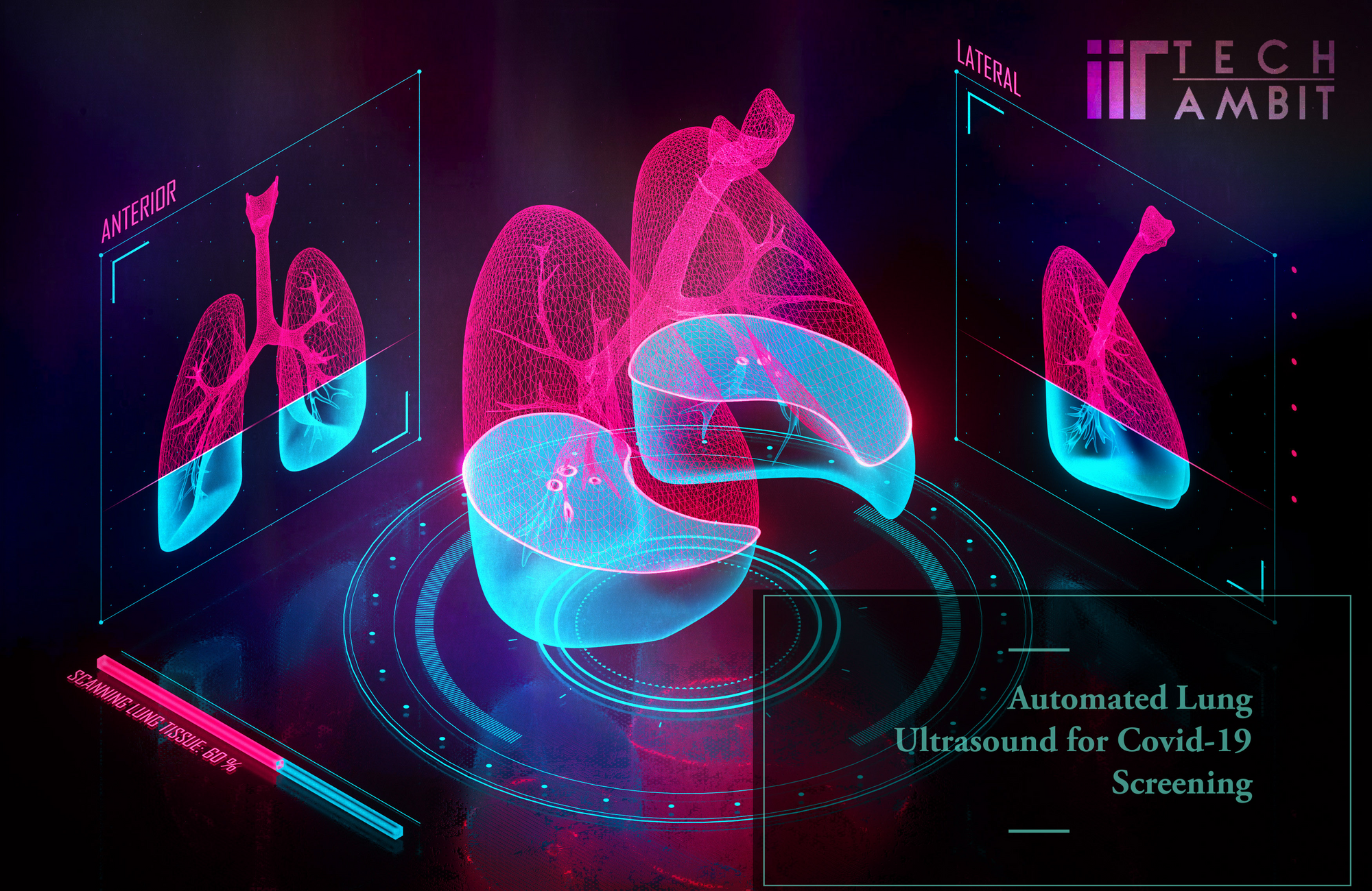
Amidst the pandemic, almost every research and development institute is looking to contribute and benefit the world. IIT Palakkad in collaboration with a few others did exactly that in the months April-June. IIT Palakkad had developed an automated lung ultrasound(LUS) for Covid-19 screening and monitoring, through a cloudbased image analysis and scoring system. The tool, which is the first of its kind in India and maybe even the world, was made available for use by clinicians to obtain an automated analysis after uploading an ultrasound video and is being constantly updated to get more accurate analyses.
Reasoning Behind the Project:
It has been reported that abnormalities in the lungs due to Covid-19 may be developed before clinical manifestations and nucleic acid detection, and due to this, it's recommended to screen suspected patients with a diagnostic imaging technique. The SARSCov-2 infection is highly contagious. The risk of transporting patients with low levels of oxygen in their blood along with blood circulation problems makes chest CT scans less viable. Lung ultrasound gives similar results as that of a chest CT scan (while being better than X-rays) with the added benefits of being easier to use at the point of care, real-time dynamic imaging, repeatability, and the absence of ionizing radiation, all while being available at a lower cost. The promise shown by the results from Italy on lung ultrasound served as a subsidiary motive for the development of this project.
The team, led by Dr. Mahesh R. Panicker started their investigation which would eventually lead them to develop the automated LUS tool. Lung ultrasound images provided by the director of the Ultrasound Division at Hospital Universitario, La Paz were used to investigate. The director, Dr. Yale Tung Chen, who had tested positive for Covid-19 shared LUS images of himself as the disease progressed. The data processing tool categorized the condition of various parts of his lungs into three: normal, viral, and bacterial infected. The conclusion is reached by first processing the images and then letting neural networks do their thing.
Functioning of the Tool:
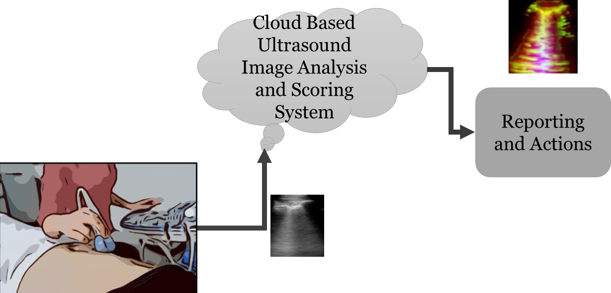
Working with Dr. Mahesh was Professor Vinod A. Prasad, and he put into words the working of the tool. The LUS analysis tool could be used by a nurse, in the absence of a skilled clinician by following a couple of steps: i) acquiring the lung images and ii) transferring the images to the cloud. These images are then analysed over the cloud and scored based on the type of infection and the severity of the infection. Professor Vinod specifically mentioned that a tool like this will be very valuable when there's a time constraint. Manually evaluating many LUS images would take quite a bit of time, but when there are multiple lives at stake, using an automated tool that is very accurate is the way to go. If the required data is provided, the tool could be extended to recognize other lung infections such as pulmonary oedema, pulmonary embolism, pneumothorax, chronic obstructive pulmonary disease and asthma. The tool also enables clinicians to study the severity of the infection. Normal lungs are depicted by A-lines, slightly infectious lungs by A+B lines, infectious lungs by B-lines or B-patches, and seriously infected lungs by consolidations.
The values pertaining to the data processing tool's results will always change based on the method of acquiring ultrasound images and the expertise of clinicians. So, it is very well understood that the tool is meant to aid a clinician in making his/her judgement, and not replace him/her. Currently, the classification of infection type has an approximate accuracy, sensitivity and specificity of 96%, 95% and 97% respectively, whereas the severity classification has accuracy, sensitivity and specificity of 97 %, 92% and 98% respectively. Currently, steps are being taken to extend the tool beyond Covid-19 detection, to act as a reliable comprehensive lung ultrasound image analysis tool during emergencies.