
“From Depth effect in dual cameras to medical imaging and 3-D printing, the evolving field of 3-D imaging might just be a game changer.”
ABOUT THE EXPERT:
Professor Chaitali Chakrabarti is a Professor at School of Electrical, Computer and Energy Engineering at Arizona State University. She has been honoured with numerous awards including Distinguished Alumni Award, Dept. of Electrical and Computer Engineering, University of Maryland, College Park (2013), Fulton Exemplar Faculty Award (2014).

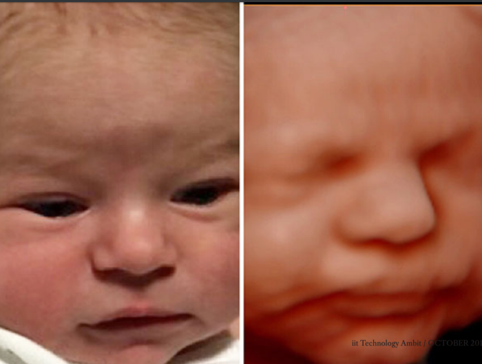
Medical imaging is the technique and process of creating visual representations of the interior of a body for clinical analysis and medical intervention. Ultrasound medical imaging is a very promising medical imaging technology because of ease of use and non-invasiveness. 3D ultrasound imaging has significantly improved accuracy in diagnosis compared to 2D imaging. However, its high computational complexity and resulting high power consumption has precluded its use in hand-held applications.
Professor Chaitali’s current work on portable medical imaging is aimed at generating high-quality 3D imaging by a small form factor device with limited power budget. 3D ultrasound imaging can also be used for accurate blood flow analysis, where the blood velocity profile is used to diagnose different types of cardiac diseases. Prof. Chaitali’s vision is to make portable 3D ultrasound imaging devices a reality. They will be available to all medical practitioners and become the ‘stethoscope of the future’.
For many years now we all have been blessed with the ability to click photos on our phone. This has pretty much revolutionized everyone’s life in the 21st century. The imaging capabilities of our phones are based on 2D imaging. How about if we could capture the world around us from all perspectives in 3D. Extensive research is going on to enable fullfledged 3D imaging on our mobile phones. While this technology is not yet available on our smartphones, it might not be a very far-sighted dream.
3D imaging relies on stereography, which we can observe from a familiar source: the human vision system. Humans see things with two eyes set slightly apart. This allows them to perceive depth in addition to the horizontal and vertical information reproduced by, for example, the standard 2D television screen. Since the eyes are separated, each one sees the world from a different angle.
The dimensionality that humans perceive in their vision comes from the brain combining disparate images into a whole – a phenomenon called parallax. Two lenses are used in every 3D shot – each captures an image slightly offset from the other. The images are edited to display while maintaining full data fidelity. The eye cannot process both sets of images on its own: each eye processes its own set of images.
3D imaging can be achieved through active or passive methods. Active systems use methods like time-of-flight, structured light, and interferometry, which generally require a high degree of control in the filming environment. Passive methods include depth from focus and light field. In snapshot-based methods, the difference between two snapshots captured at the same time is used to calculate the distance to objects – this is called passive stereo imaging. It can be achieved by moving a single camera, but using two cameras with identical specifications is more efficient.
3D imaging has opened possibilities for enhancement and new innovations in a plethora of avenues. We can already see all the latest smartphones coming with dual camera, where the dual camera uses 3D imaging to create that depth of field effect. Several companies have developed apps which are capable of 3D modeling at a very moderate level.
Experiments have been going on with augmented reality and 3D image manipulation. These will allow users to play with 3D images that can be created using a 3D printer, enabling a more interactive user experience. Archeologists are also leveraging 3D imaging technology to help preserve sites and findings. Because excavating dig sites creates a high risk of destroying the story behind it, archeological professionals are eagerly turning to 3D modeling technology in order to help preserve the information found in a site, discover new areas to start digs, and to create models of ancient artifacts using 3D printing.
In the medical front, prosthetic limbs can be manufactured at a much lower price with advancement in 3D imaging. Facial implants can be done much easily with reduced risk. Diagnosis of heart diseases can be enhanced by building an extremely accurate real time 3D anatomic model of patient’s heart which will improve diagnosis and treatment.
3D imaging also allows for the creation of highly-detailed medical models, giving medical practitioners an inexpensive alternative to learn about the human anatomy without having to use actual human body parts.
We can but predict how the amazing future prospects of 3-D imaging will lead to enhancement in a myriad of sectors. We can be excited and optimistic about how of 3-D imaging will morph our near future!