

Background
Caused by a mosquito-borne virus, Dengue is a serious global health concern in the current era. It is prevalent in tropical and sub-tropical areas like southeast Asia, America and western pacific regions. Early diagnosis of such diseases is essential to provide timely management to the patient and prevent health deterioration to severe illness.
However, existing diagnostic methods like virus isolation, nucleic acid detection are time-taking (may even take a week), require a lot of expensive equipment and reagents and need technical expertise, limiting their use for early detection. The need of the hour is a rapid, inexpensive, user-friendly on-site diagnosis that requires minimal infrastructure and is scalable to the community level.
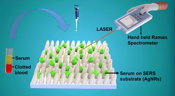
The technique presented by the GLAD research group at IIT Delhi’s Physics Department meets most of these requirements. They have created a Surface Enhanced Raman Spectroscopy (SERS) based Platform that gives dengue tests results within an hour.
The research work which was published in The Journal of Analytical Chemistry and Journal Colloids and Surfaces B: Biointerfaces, was funded by Program IMPRINT INDIA, a joint initiative of Indian Institute of Technologies (IITs) and Indian Institute of Science (IISCs) seeking to develop road map for research to solve major engineering and technological challenges.
When asked about the inspiration and the series of events that led to their exemplary work, The project’s principal investigator, J.P. Singh, Professor at IIT Delhi recalled it was during his sabbatical at the university of Georgia that he was observing the raman spectra of Hendra virus measured by department of infectious diseases where he developed a keen insight into the idea.
“I got the idea like why not? Although the setup they had back there was a huge benchtop raman setup, I realized we could also use this idea along with the sensors we were using at the point. And so, after coming back, we started discussing it within our group which has been working on GLAD (Glancing Angle Deposition) for 15 years.”
The main idea behind the set-up is the well-known technique of Raman Spectroscopy.
Raman Spectroscopy
A sample scatters only a tiny part of light falling on it, about 1 %. 99% of this scattered light consists of photons of the same frequency as the incident light known as Rayleigh-scattering. The rest of it is shifted in both wavelength and energy, known as Raman-scattered light.
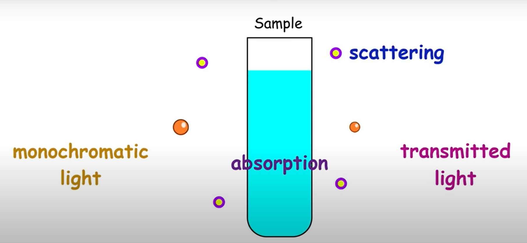
The spectrum of the Raman-scattered light depends on the molecular constituents of the sample and hence is specific to that material. That makes it helpful to identify any substance (including biological materials) by its Raman spectrum. In fact, they are known as molecular fingerprints for this reason.
A popular method for detecting viruses in the body is to check for the presence of their antigens. An antigen is a toxin or other substance given off by a virus that causes an immune response in our body. Raman Spectroscopy serves as a great tool to detect and identify these antigens. For example, the presence of NS1 antigen confirms Dengue.
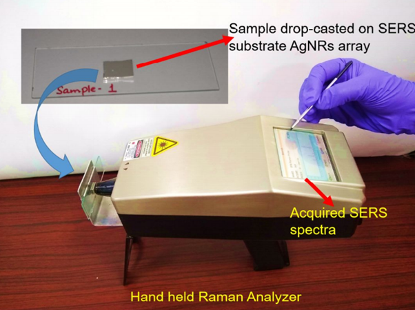
This can be quickly done on-site at a hospital/institute with the help of a handheld Raman spectrometer. It shoots a laser beam at the sample, collects the scattered light and displays the resulting spectrum (intensity – wavelength graph).
However, biological materials scatter light poorly, further reducing the already low scattered light intensity, leading to weak Raman signals that make it difficult to detect the antigen at lower concentrations.
SERS Substrate
To tackle this problem, , the research group headed by Principal Investigator Professor J.P. Singh used a technique called GLAD to fabricate thin roughened up metal surfaces (usually made of noble metals like Gold) with different shapes, built with precision up to the nanometer range.
Then, silver nanoparticles (having a diameter between 1 to 100 nm) from a solution were deposited on the metal surface.
The resulting metal surface, known as a SERS platform, acts as an amplifier, i.e. when shone on with a laser, the surface electrons on these metal structures get excited and create an enhanced electric field localized near the surface.
The degree of enhancement greatly depends on the dimensions and shape of the SERS platform and the nature of the nanoparticles used. However, by utilizing the optimum combination, the researchers obtained electric fields as high as 108 times the original one.
When the dengue samples to be tested were adsorbed on the platform, and their spectra analyzed, the Raman signals obtained were enhanced by the same factor!
With this method, spectral data can be obtained even at low concentrations of the antigen. This increases the specificity (proportion of true negatives) of the diagnostic test by eliminating false negatives. The use of SERS would tremendously increase the application of Raman Spectroscopy in medicine and allied fields.
Testing
The handheld device has been successfully tested on clinical blood samples collected from a fever clinic at the Indian Council Of Medical Research (ICMR) – NIMR, New Delhi.
Blood samples were taken from fever patients as well as healthy individuals, and the spectral data obtained was fed into a Principal Component Analyzer (PCA) that is a statistical tool for data analysis. Based on the input data, It accurately differentiated between the three sets of blood samples, Dengue positive, Dengue negative and healthy patients for any samples tested from thereon.
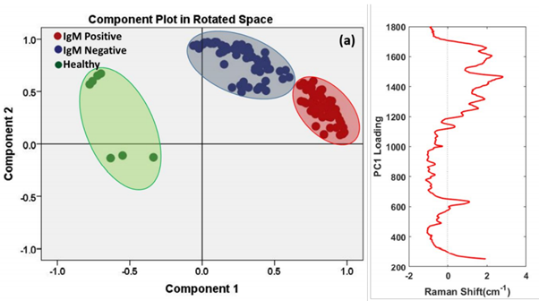
“Getting spectral differences manually by just looking at the spectra is error prone and less reliable. Sometimes if you are lucky, you might get all the peaks within the spectra. But this is usually difficult for most of the viruses. Hence, we had to resort to Statistical tools like Principal component analysis to feed the spectral data for separating the different classes. (PCA works by reducing the dimensionality of datasets, thus increasing interpretability while minimizing loss of information)”
Explained Prof J.P. Singh, talking about the importance of data statistical tools such as PCA (Principal Component Analyzer) for differentiating samples
The SERS technique is not only limited to Dengue and can be extended to detect a host of other viruses and bacteria. For example, in collaboration with the ICMR- National Aids Research Institute (NARI), the research group successfully demonstrated the detection of the human immunodeficiency virus (HIV-1) through this handheld SERS platform.
Prof Singh also talked about the potential range of applications of the technique in other domains,
“People are increasingly utilizing such ideas for detecting explosives and for differentiating toxic chemicals since that becomes much quicker and easier through SERS with the molecules having different functional groups attached with them.”
When asked about the hardest aspect of the research, Prof Singh explained
“The problem mainly lies with the kind of pathogen which we are looking for. To give an insight, suppose you wish to separate and distinguish chemicals using the spectra from SERS. Now this becomes relatively easy due to the fact that the respective chemicals have two separate functional groups, hence their ‘structural signatures’ are different which can be interpreted from their obtained spectra. However, the case with biological pathogens and viruses becomes complex due to the fact that their outer envelope is composed of proteins which are more or less similar in nature hence resulting in only minor changes in the spectra. The fact that there are frequent mutations occurring at the cellular level doesn’t help either”
Another problem posed is due to the contaminants. Since the Enhancement factor in Surface Enhanced Raman Spectroscopy (SERS) is high, the increased sensitivity leads to enhancement of net signal intensity. While this does facilitate analysis of spectral data at lower concentrations of antigen, this also makes spectral data prone to alterations by small quantities of contaminants.
Conclusion
Discussing the bigger picture with respect to the need for rapid and sensitive testing techniques, Prof J.P. Singh said
“Although some of the rapid diagnostic kits prevalent currently are very useful and are able to provide quick results, one vital problem associated with them is of specificity. Such techniques are prone to false positives and false negatives. The SERS based technique is highly specific and quick enough to produce the final report of investigation within an hour once you’ve made the reference library for the concerned sample and the intervention of skilled labourers is much less once the program is trained. There’s still a lot of potential and room for improvement. But in the future I would like to see more specific and accurate sensors and techniques which are based on less-complex ideas.”
It may seem that sophisticated fabrication methods drive up the expenses, however, a SERS chip developed by this technique costs no more than a dollar, and the Raman spectrometer makes for a one-time investment by the hospital/institute. In all, the SERS platform shows excellent potential to replace the traditional diagnostic methods used nowadays.