There’s loads of things one can make in a lab. Organic compounds, mayhem, brains? One of the biggest problems with neuroscience research is how well protected human brains are. Tucked away within a chamber made of 22 bones, surrounded by 3 membranes to cushion against shocks, our brains are effectively locked up in our bodies’ highest security vault. While this is great for protection in everyday life, this is incredibly frustrating if one is trying to study said brains and needs to observe them at work or to extract them safely.

And while studies have been done on animals’ brains, they differ so vastly from those of humans that they did not offer much insight, once the basic working was established. Autopsies and digital imaging techniques have also been used for studies and while they have contributed a lot to our understanding of neuroscience, we still do not comprehend how most disorders (e.g. Alzheimer’s) work. As most deviations and aberrations appear during the developmental stage of the brain, it is absolutely essential to be able to observe the brain while it was developing. Here’s where cerebral organoids come into the picture.
What are cerebral organoids?
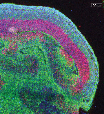
A brain or cerebral organoid is a self-organizing 3-D tissue which is able to simulate the architecture and functionality of the human brain.
How are they made?
Researchers can obtain human neural cells by differentiating human pluripotent stem cells (cells that are able to self-renew by dividing and developing into groups of cells that make up a human body).

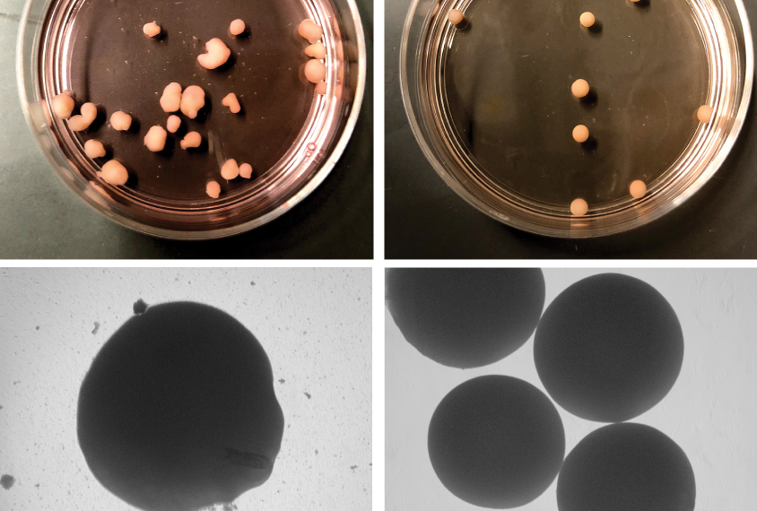
The biggest challenge faced in the discovery of making these cerebral organoids was coming up with the perfect combination of nutrients to induce the stem cell to develop a neural identity, which was finally discovered in 2013. The rest of the process is relatively simple, as the organoid basically grows itself, in the presence of this combination of proteins, vitamins, minerals and sugars, a special gel (to simulate embryonic tissue), a warm incubator (at body temperature) and some motion (to mimic blood flow). The stem cell grows into a mini-version of an early developing brain. It can form simplified neural networks and closely follows the stages of fetal development.
How do they differ from an actual brain?
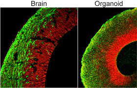
A cerebral organoid is made up of the same types of tissues as a real brain, however, they are organized quite differently. Hence, they are as different from an actual brain as a Lego house is from a Lego plane.
Also, these simplistic models are tiny in comparison with real human brains. A full-sized human brain has 86 billion neurons on average, against the 100,000 neurons in an organoid. And since brain organoids don’t have blood vessels to feed them, their size is very limited, generally a few millimeters in diameter.
One must also take into account the neural pathways built by the human brain by interacting with its surroundings. Since the organoids are essentially isolated, disassociated brains, they can’t interact with their surroundings. So, while useful similarities exist, the differences are significant and organoids aren’t simply smaller versions of real brains.
Applications
Cerebral organoids have the potential to function as a model to study mental diseases and disorders. However, diagnostic tools are needed to evaluate the tissue and create organoids that mimic the disease or state of development in question. Other potential uses include cell fate potential (determination of how a particular cell develops into a final type) and cell replacement therapy. They also offer insight into the timing and order of neural development in animals and can even be used to study cell migration. Basically, they're that cousin to the other models (you know the annoying one who is the class topper and the best sportsperson ever and can somehow also paint, sing, and dance?)
They can also serve as preliminary tests for drugs, to check for efficacy as well as safety. For instance, studies on organoids helped determine the chief enzymes responsible for the effects of cocaine as a teratogen (birth defect causant), one of them being cytochrome P450 isoform CYP3A5.
Studies on diseases
>>> Microcephaly: is a developmental condition in which the brain remains undersized leading to an undersized head. Its study was not suitable for mouse models, but organoids helped formulate theories about the condition which suggest the underlying mechanism to be a span of overly rapid development, which causes a deficit of glial cells and an eventual reduction in the rate of brain growth.
>>>Alzheimer's: Organoids from PSC’s of affected patients were modelled against those from those of healthy individuals. The affected models were found to have structures similar to that of plaques caused by amyloid beta proteins and neurofibrillary tangles, which cause the disease's symptoms.
>>> Autism spectrum diseases: Cerebral organoids from ASD macrocephaly patients were found to reflect characteristics typical of the ASD-related macrocephaly phenotype found in the patients, which led to previously undiscovered connections between certain gene mutations and phenotypic expression. Autism has also been studied by the healthy versus affected organoid model, which showed an overexpression of a transcription factor FOXG1 that produced a larger amount of GABAergic inhibitory neurons in the affected models. The significance of this use of brain organoids is that it has added great support for the excitatory/inhibitory imbalance hypothesis which if proven true could help identify targets for drugs so that the disease could be treated.
>>>ZIKA: The teratogenic effects of the Zika virus, with many cases leading to microcephaly (having an abnormally small head for a particular developmental stage) were studied using organoids. The effected organoids were smaller than their healthy counterparts, had increased apoptosis (body-regulated cell degeneration and death), reduced NPC (neural progenitor cell) populations and increased lumen size. Also, when organoids at different stages of development were exposed to the Zika virus, studies found that earlier exposures had the most disastrous effects.
Limitations
Cerebral organoids make preferred models because they reflect the structure of the human brain rather well and follow through the stages of fetal neural development for an extended period of time. Their potential is immense! However, there are a few factors which prevent them from being the neuroscience equivalent of the number 42 (yes, that was a hitchhikers reference.) For instance, it takes several months to create an organoid and analyze it. Also, since they do not have many of the structures typical of a human brain, it limits the types of diseases that can be studied. Central parts of the organoid tend to start dying out due to lack of oxygen and nutrients. However, this can be avoided by using fluidic devices and hence, increasing exposure to media. The structure of cerebral organoids can be variable, due to differences in stem cells used as well as different manufacturing methods. Also, their maturation is limited to about the normal developmental level of the second trimester, so they can’t offer any information about late-onset disorders.
A lot of people think that the initial step that we have made, of just developing the organoids in the first place, was the really big jump. I would of course like to think so because I had a major part in that, but I actually think it is much harder to get blood vessels in there, and get it to develop beyond where it currently is.
Neurobiologist Madeline Lancaster
Ethical questions
Growing cerebral organoids in labs gives rise to a multitude of moral questions like: Can they develop a consciousness? Can they think for themselves? Can they feel pain? Presently, this seems highly unlikely, given how simple the organoid models are.
Well, what would they be conscious of?
Christoph Koch, American neuroscientist
However, they are shown to give responses to light-based stimuli. If proven, the affirmative of any of these questions would raise grave moral issues and are the subject of much debate and discussion.
Future
Brain organoids have currently been around for less than a decade and their research is still in its infancy. Their limited maturation places rather rigid boundaries on their usefulness and this arises mainly from the lack of a cardio-vascular system. However, in the future, brain organoids could be combined together with blood vessel organoids to study neurovascular interactions and to support long-term culture, which would give unprecedented insights into late-onset diseases, stages of further development of the brain etc.