
OCT is an Emerging Technology for Biomedical Imaging and Optical Biopsy giving us the ability to dive deeper within into the layers of the retina and understand the complexities with ease using state-of-the-art Machine Learning techniques.

We all know about this complex engineered visual receptor organ known as ‘The Eye’. With what ease it distinguishes & classifies different objects contrasting to a state of the art deep neural network algorithm or computer vision software. But do you know sensory intellect comes at a cost? A cost where you can lose this visual prowess forever! And you might not be even aware of it.
With age and time, our Eyes and the retina starts to lose its integrity and the ability to interpret signals from the visual cortex of the brain thus pacifying and fading into oblivion. A big reason is being unaware of what happens within. The vitreous humor that holds all the layers in place by applying force on them, starts to lose its viscosity. Subsequently, the retinal layers that were tightly bonded and communicating at a phenomenal rate are suddenly put into a jolt. A jolt that makes them loose from each other and breaking of micro neurons. But this is not where the problem begins. Our body knows how to tackle this by adjusting the cornea and the lens. The real danger lies in the formation of a tumor in the choroid layers with prolonging inattention to the eyes. This tumor is capable of hampering your eyesight forever.
I’ve told you about where the problem lies but I haven’t told you about why we can’t rectify this problem with the ease we solve other problems. Firstly, it's very arduous and impossible to predict the tumor size and location by seeing the external eye. We need to dig even deeper and this is where Optical Coherence Tomography kicks in. Optical Coherence Tomography or OCT is the technique that uses a laser projection to map the internal surface of the eye. The laser enters the eye and hits the retina causing a spike in the frequency of the laser. This spike is recorded on a graph and using the correlation to the thickness of retinal layers, the geometry is plotted.

To understand more on this complex subject we go to Divyadrishti Imaging Laboratory at IIT Roorkee. The main theme of the research Lab is creating hardware and post-processing soft tools, mainly, related to Direct and Non-Invasive Imaging for Industrial and Medical Applications. Non-Destructive Evaluation is also one similar field. The Major objective of research is to pose challenges so as to improve the understanding of Radiation Propagation and its measurement, in general.
The team from Divyadrishti Imaging Laboratory, IIT Roorkee tells us, "There are only 3-4 OCT machines on the Earth! With one of them being at IIT Roorkee. Our mission is to provide better healthcare facilities in the domain of optometry."
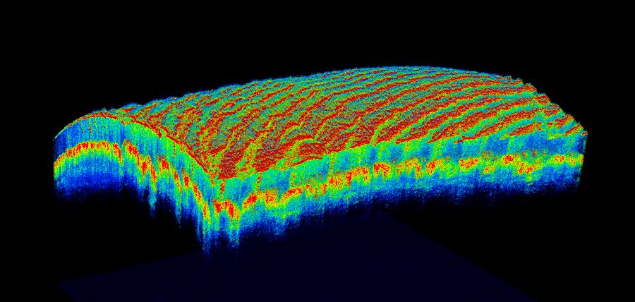
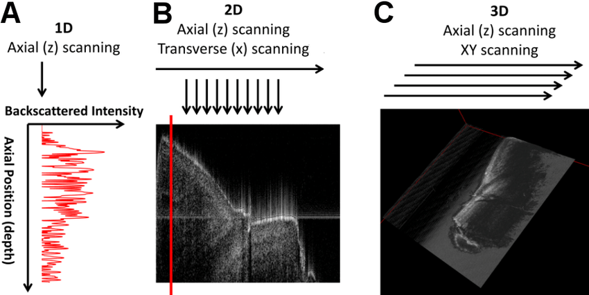
The team also tells us that there are three types of OCT scans namely — OCT-A, OCT-B & OCT-C. The major differentiation factor among the three is the dimension of the scan. In OCT many one-dimensional scans (A-scans) are performed at several depths to create a two-dimensional image (B-scan). Those B-scans, if acquired closely and rapidly, can be translated into a volumetric image (C-scan) of a retina,
So we do have a possible solution to take a glimpse into the internal framework of the eye. So why can’t we rectify this tumor? Our modern-day techniques only allow us to take an image of the internal working of the eye but don't classify whether it has a tumor or not. This is where doctors scratch their heads as they cannot tell if the retina has a tumor or not with exact assurance. There might be a lot of false-positives that may lead to unnecessary treatment or a lot of false-negatives leading to failure of treatment. So what is the one true solution?
Deep Learning! This is where the solution lies. I may have said in the beginning that The Eye surpasses every possible deep learning model. So how can it detect the tumor? The secret lies in the capability of a model learning the same thing for a billion years without complaining. It’s not a human that grows weary of doing the same task for more than a day. That’s what truly makes it a machine, right? A neural network has the capability to see several images of an OCT scan and learn how a healthy eye differs from an infected one. It becomes so capable that it can differentiate a tumor from a normal eye on a single pixel basis! A doctor might improperly classify a fluid pocket in the retina or loose retinal layers as a tumor but a neural network will not.
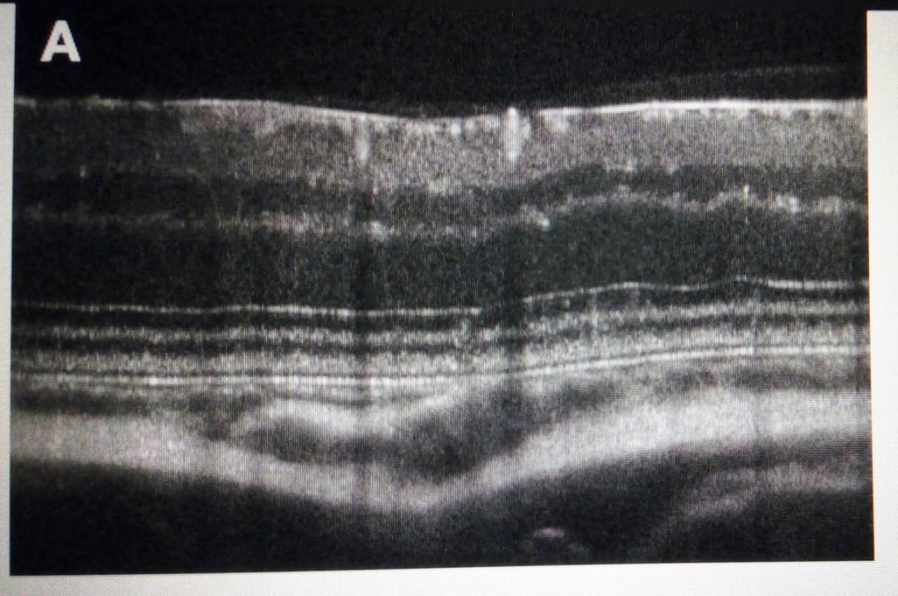
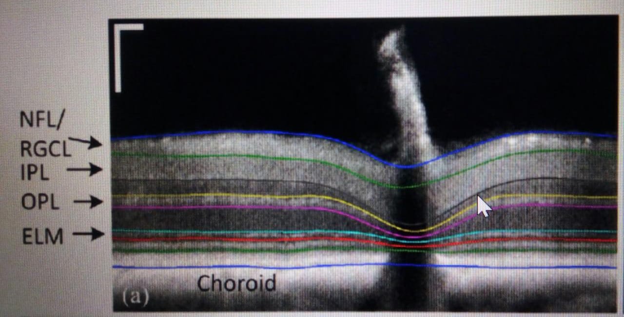
Preprocessing: The process begins by taking an OCT-B scan as shown above and breaking it into several layers namely, photoreceptors, neurons, and choroid. This is a challenging task as there is a lot of noise in the data and manual annotation of data is tiresome. A few attempts are being made to make this process automated.
One such attempt is being done by Divyadrishti Lab at IIT Roorkee. The team has used several convolution neural networks to self annotate the image and extract all features and artifacts from it.
The automation of this process will lead to a revolutionary change in the learning process by decreasing the time taken 100 folds. After the data is annotated, it is converted into the desired format as the training model demands. It can in the form of pixel values or greyscale values.
Training Phase: The real task of making your model learn begins! The data is labeled as healthy and infected and fed to the model. The model like a naïve child starts learning the true difference at single-pixel levels. It is made to learn the data repeatedly and then used to predict unseen OCT scans. The process is truly magical if looked through the perspective of the network. The whole process is done using various libraries as Scikit-Learn and Tensor Flow which are toolkits to work on machine learning projects. We don't need to worry about the internal working about the model right now. What we really want is our model to predict with maximum true positives.
"It doesn't matter what algorithm or toolkit you use. The most important task is to structure your learning towards detecting the retinal layers, even though the accuracy is low." - Team at Divyadrishti Lab, IIT Roorkee
Deployment Phase: After the desired accuracy is attained the model is directly implemented into an OCT graphing machine. The machine automatically tells whether the scan has a tumor or not with a definite quantifiable assurance. This leads to a better-secured approach to this difficult task of identifying the tumor.
The Big-AI dream has always been about automating everything. But its heart truly lies at the intensive problems as identifying a tumor. What may seem simple at first has several layers of complexities attached to it. Solving such a problem truly enlightens us about this complex structure of healthcare. The aim of mankind has always been to be better at what it does. And AI is nothing but an ally to us at what we do!