
The omnipresence of electronics today can be attributed to a series of inventions, starting mainly from the transistor. These inventions helped reduce the size of devices and improve their performance by a huge factor. As electronic circuits were made smaller, their components got faster, required less power, and became cheaper to produce. Thinking along similar lines, muscope was created.
The motivation behind Muscope
Optical microscopy is indispensable to life sciences and biomedical applications. Conventional light microscopes remain costly, complex, bulky, fragile, and largely manual. A significant yet generally overlooked shortcoming of these microscopes is their inflexibility towards integration into bigger systems, which is an obstacle for a whole class of new devices and automation.
Several palmtop-sized mini-microscopes also do exist. All these designs feature the removal of the lens train. To improve the resolution, usually, a light source like a laser is used, which is not easy to minimize and operate. The employment of macroscopic illumination systems limits their scalability.
Developed by Ekta Prajapati, Saurav Kumar and Shishir Kumar, muscope aims to fix the above issues. It extends only a few mm in each direction and is capable of high-resolution imaging.
How Muscope works
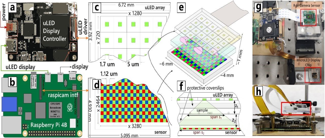
Muscope achieves these feats by replacing the extended light source with state-of-the-art micro LED displays. Micro LEDs are micron-size LEDs available in various colors. The underlying technology allows high brightness so that even a single micro LED is adequate for imaging.
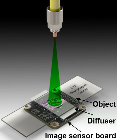
Broadly, muscope comprises two electronic chips- image sensor chip and micro LED display chips. A driver board interfaces micro led display with a single board computer. The display of muscope consists of micro LEDs arranged on a rectangular array. The micro LEDs can be individually switched on, and they are very bright.
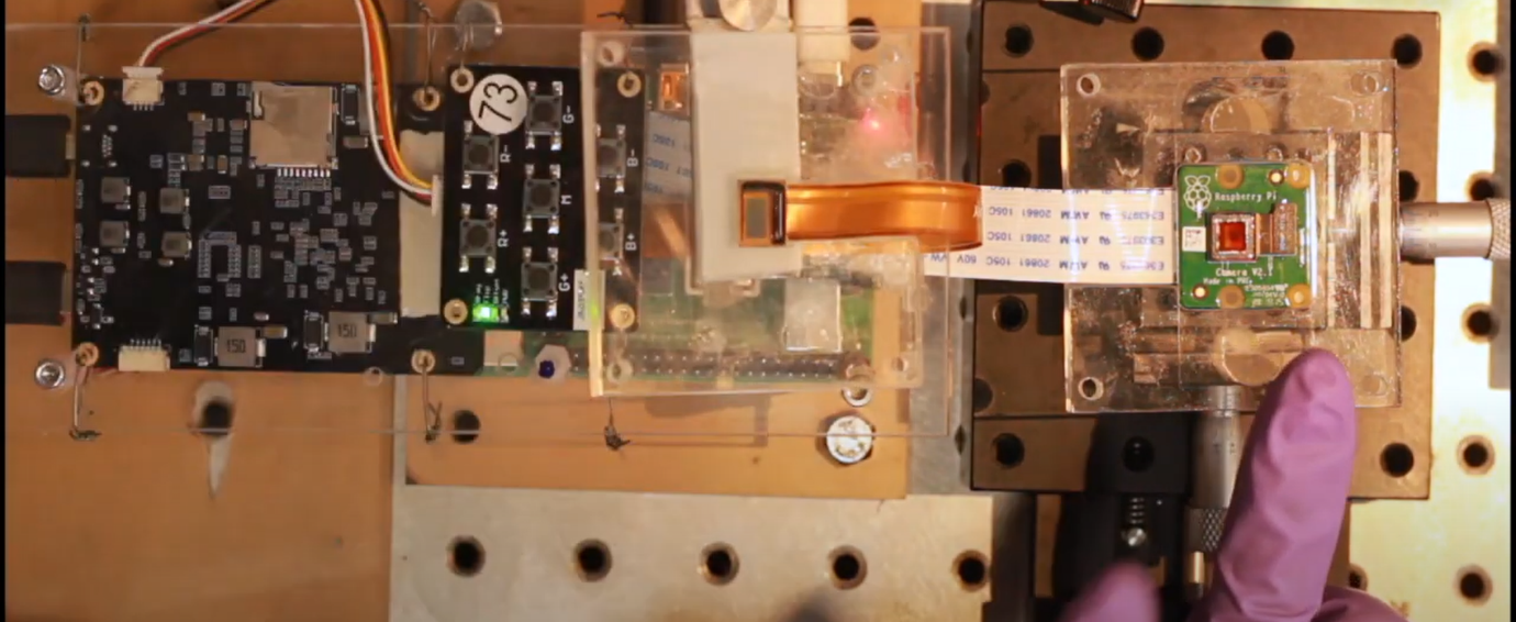
The micro LED control board is used to control the brightness and actuation of micro led chips. The sample is placed between the micro LED chip and the image sensor chip. To perform imaging, it is required to synchronize the illumination of the micro led chip and the camera sensor chip using a python script. Muscope is lens-less. Therefore, the sensor captures holograms, from which real images are reconstructed.
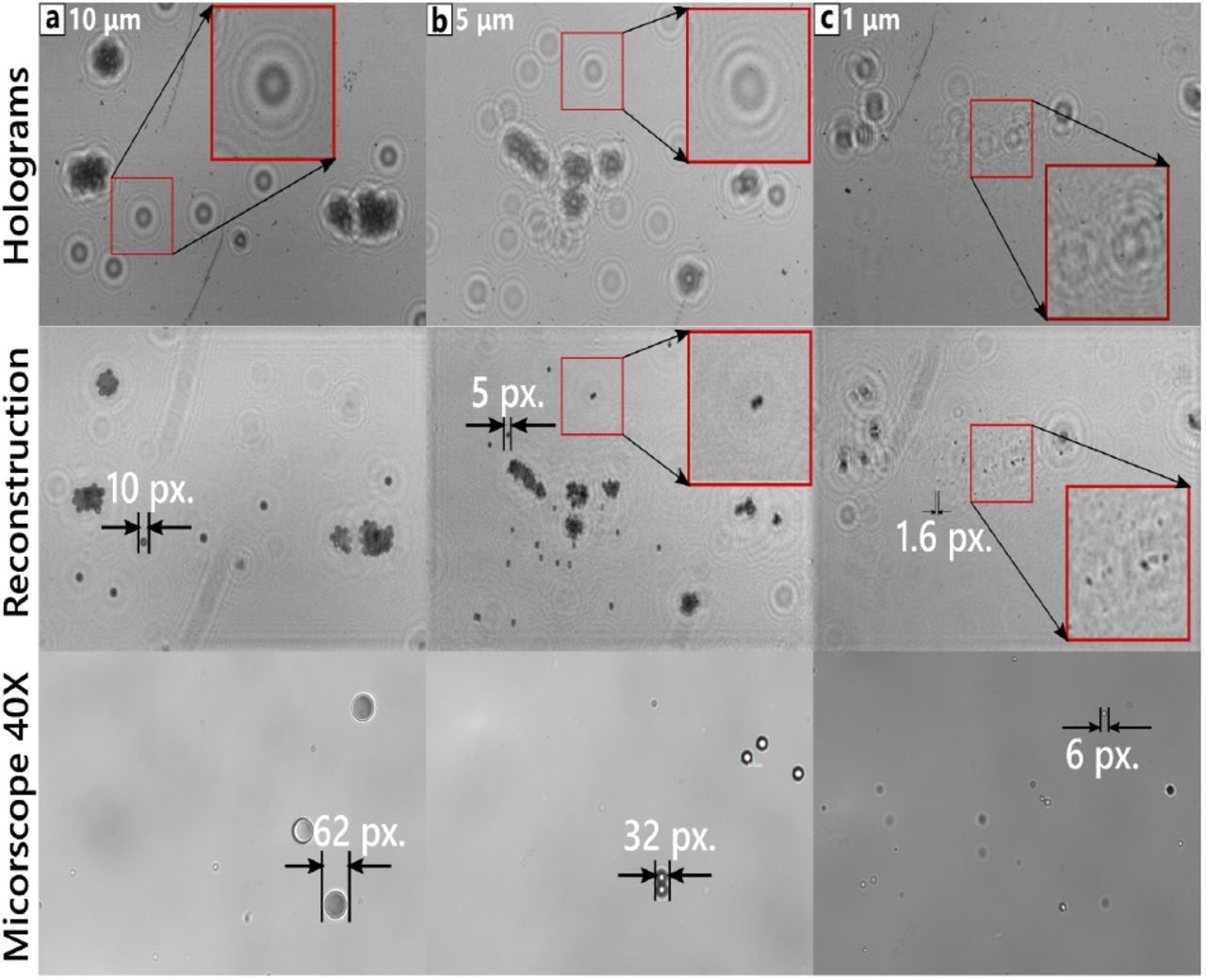
Micro-scale movement of light source by electronic means is perhaps the most salient feature of Muscope, which allows implementation of software-controlled super-resolution. The illuminating light coming from a micro LED can be moved around in a two-dimensional plane by selecting a different micro LED from the array.
The movement of light sources makes it possible to capture images of widely separated regions of the sample, effectively expanding the field of view (FOV). This movement can be used for capturing images of samples under slightly different lighting conditions. Such images can then be stitched to increase the resolution.
Applications
Muscope will make medical devices low cost, improve their mobility and automation. Resource-constrained or specialized medical devices, extreme conditions of environmental monitoring, agriculture, and animal husbandry are domains where Muscope can excel.
For example, muscope can be integrated with microfluidic chips. This allows manipulation of single cells in these chips, dosing them with chemicals, and observing them clearly under various conditions. In fact, it can be hermetically sealed in a microfluidic device such that the channel lies between the micro LED and the image sensor chip.
Enhancements and further
Muscope renders multiple future possibilities, mainly due to its compatibility with microfluidic technology and robotic technology. It can be scaled down further from its already tiny form to harness the benefits of deep integration, enhanced mobility, lower cost, and robustness.
The researchers are keen on commercializing the technology, expanding the work to disease detection using biomarkers, miniature analysis systems, and machine learning-aided workflows.